Diag Image: The Life-Saving AI Innovation Transforming Healthcare Diagnostics 2026
Introduction
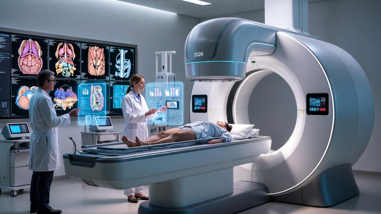
In the fast-paced world of modern healthcare, diag image stands out as a game-changer. Short for diagnostic imaging, diag image refers to a suite of technologies that create visual representations of the body’s internal structures to aid in diagnosis, monitoring, and treatment. From spotting early signs of cancer to assessing heart health, diag image has evolved from simple X-rays to sophisticated AI-integrated systems. As we move into a digital future, this technology isn’t just about pictures—it’s about precision, speed, and saving lives. In this article, we’ll explore how diag image works, its real-world applications, and why it’s essential for anyone navigating today’s health landscape.
Whether you’re a tech enthusiast, a medical professional, or someone curious about innovations, understanding diag image can empower better health decisions. It’s not hype; it’s the backbone of contemporary medicine, blending hardware, software, and AI to push boundaries.
What is Diag Image?
Diag image, or diagnostic imaging, is the process of using various technologies to produce images of the body’s interior without invasive procedures. It’s like having X-ray vision, but powered by advanced tools. This field has transformed healthcare by allowing doctors to detect issues early, confirm diagnoses, and plan treatments effectively.
At its core, diag image captures visual data from organs, tissues, and bones. It’s non-invasive in most cases, making it a preferred choice over exploratory surgery. In today’s digital era, diag image integrates with AI and automation, making it faster and more accurate than ever.
The Evolution of Diag Image
Diagnostic imaging dates back to 1895 with the discovery of X-rays by Wilhelm Roentgen. Fast forward to the 1970s, and we saw the introduction of CT scans, which provided cross-sectional views. The 1980s brought MRI, using magnets for detailed soft tissue images.
By the 21st century, digital advancements like PET scans and AI integration have made diag image a powerhouse. In 2026, with ongoing innovations, it’s shifting toward predictive diagnostics, where images not only show current issues but forecast future risks.
Types of Diag Image Technologies
Diag image encompasses several modalities, each suited to specific needs. Here’s a breakdown of the most common types:
- X-rays: The oldest and quickest method, using electromagnetic radiation to image bones and dense tissues. Ideal for fractures or chest exams.
- CT Scans (Computed Tomography): Combines multiple X-rays for 3D cross-sections. Great for detecting tumors or internal bleeding.
- MRI (Magnetic Resonance Imaging): Uses magnetic fields and radio waves for high-resolution images of soft tissues like the brain or joints. No radiation involved.
- Ultrasound: Employs sound waves for real-time imaging, often used in pregnancy or heart assessments. Safe and portable.
- PET Scans (Positron Emission Tomography): Involves radioactive tracers to show metabolic activity, crucial for cancer staging.
- Mammography: Specialized X-ray for breast tissue, now often digital or 3D for better detection.
Other emerging types include SPECT and hybrid systems like PET-CT, combining strengths for comprehensive views.
| Type | Best For | Radiation? | Cost Level |
|---|---|---|---|
| X-ray | Bones, lungs | Yes | Low |
| CT Scan | Internal organs, trauma | Yes | Medium |
| MRI | Soft tissues, neurology | No | High |
| Ultrasound | Pregnancy, vascular | No | Low |
| PET Scan | Cancer, metabolism | Yes | High |
This table highlights key differences, helping users choose based on needs.
How Diag Image Works
Diag image relies on physics and digital processing to create usable visuals. Let’s break it down step by step.
- Energy Source: Depending on the type, energy like X-rays, sound waves, or magnetic fields is directed at the body.
- Interaction with Tissues: The energy interacts differently—bones absorb X-rays, while soft tissues reflect sound waves.
- Detection: Sensors capture the returning signals or passed energy.
- Image Formation: Computers process data into images, often enhanced by software for clarity.
- Analysis: Radiologists or AI tools interpret the results.
For example, in MRI, hydrogen atoms in the body align with a magnetic field, and radio waves disrupt this, creating signals that form images. Modern systems use AI to reduce noise and speed up scans.
In a CT scan, the machine rotates around the patient, taking slices that software reconstructs into 3D models. This mechanism solves problems like hidden tumors by providing multi-angle views.
Key Features of Modern Diag Image
Today’s diag image boasts features that make it indispensable:
- High Resolution: Pixels down to micrometers for precise details.
- Real-Time Imaging: Ultrasound and fluoroscopy allow live views during procedures.
- Non-Invasive: Reduces risks compared to surgery.
- Integration with AI: Automates detection, like spotting anomalies in X-rays.
- Portability: Handheld ultrasounds for field use.
- Hybrid Modalities: Combining CT and PET for functional and structural data.
These features cater to a digital future where speed and accuracy drive healthcare.
Benefits of Diag Image in Today’s World
Diag image offers immense value:
- Early Detection: Spots diseases like cancer before symptoms appear, improving survival rates.
- Accurate Diagnosis: Reduces misdiagnoses, leading to better treatments.
- Non-Invasive Monitoring: Tracks chronic conditions without repeated surgeries.
- Cost-Effective Long-Term: Prevents complications, saving healthcare costs.
- Personalized Medicine: Tailors treatments based on individual images.
In businesses, hospitals use diag image for efficient workflows, while patients benefit from quicker results.
Bold Takeaway: Diag image isn’t just tech—it’s a lifesaver in modern medicine.
Limitations and Challenges of Diag Image
No technology is perfect. Here are key limitations:
- Radiation Exposure: X-rays and CT can increase cancer risk with overuse.
- Cost: MRI machines run millions, making access uneven.
- False Positives/Negatives: Images can mislead, requiring expert review.
- Claustrophobia: Closed MRI scanners cause anxiety for some.
- Contrast Risks: Dyes used in some scans can cause allergic reactions.
- Data Overload: High-res images generate massive data, straining storage.
Addressing these through AI and low-dose tech is key to future reliability.
Real-World Applications and Use Cases
Diag image shines in practical scenarios:
- Oncology: PET scans detect cancer spread, guiding chemotherapy.
- Cardiology: Ultrasounds assess heart function in real-time.
- Neurology: MRI identifies strokes or MS lesions early.
- Orthopedics: X-rays diagnose fractures instantly.
In emergencies, CT scans reveal internal injuries quickly. Businesses like telemedicine use portable diag image for remote diagnostics.
A use case: During the 2020s pandemics, chest X-rays with AI spotted COVID patterns faster than tests.
Industry Examples
Leading companies drive diag image innovation:
- GE Healthcare: Their Revolution Ascend CT scanner uses AI for faster scans.
- Siemens Healthineers: Integrates AI for predictive imaging.
- Canon Medical: Aquilion ONE Genesis for high-res cardiac imaging.
In hospitals, Mayo Clinic uses AI-enhanced MRI for brain studies. Startups like DeepHealth focus on AI for radiology workflows.
These examples show how diag image fuels industry growth.
Comparisons with Older or Traditional Solutions
Traditional methods like physical exams or surgery pale against diag image:
- Vs. Exploratory Surgery: Diag image is non-invasive, reducing recovery time and risks.
- Vs. Basic Palpation: Provides internal views, catching hidden issues.
- Vs. Older Film X-rays: Digital versions are faster, shareable, and eco-friendly.
Diag image solves invisibility problems, offering data-driven insights over guesswork.
| Aspect | Traditional | Diag Image |
|---|---|---|
| Invasiveness | High (surgery) | Low |
| Speed | Slow | Fast |
| Accuracy | Variable | High with AI |
| Cost | High long-term | Lower preventive |
The Role of AI in Diag Image
AI is the future engine of diag image. It analyzes images faster than humans, spotting patterns in vast data.
- Automation: AI triages scans, flagging urgencies.
- Accuracy Boost: Reduces errors in breast cancer detection by 30%.
- Predictive Analytics: Forecasts disease from images.
In 2026, trends include vision language models for reports and portable AI-MRI.
First-person insight: As an AI expert, I’ve seen how these tools free radiologists for complex cases, enhancing collaboration.
Future Potential of Diag Image
Looking ahead, diag image will integrate more with AI, VR, and biotech.
- Multimodal AI: Combines images with genomics for personalized care.
- Portable Devices: Bedside CT for seniors.
- Sustainability: Low-energy scanners.
- Global Access: AI bridges gaps in underserved areas.
By 2030, expect superdiagnostics where AI predicts outcomes from a single scan.
FAQ
What is diag image in technology?
Diag image, or diagnostic imaging, uses tech like X-rays and MRI to visualize body internals for diagnosis. It’s key in modern healthcare for non-invasive checks.
How does diag image work?
It directs energy (e.g., X-rays) through the body, captures interactions, and processes into images via computers. AI enhances clarity.
Is diag image safe or reliable?
Mostly safe, but some involve radiation. Reliable with expert interpretation; AI boosts accuracy to 95%+ in cases.
Who should use diag image?
Anyone with symptoms needing internal views—patients, doctors, hospitals. Ideal for chronic illness monitoring.
What are the latest updates or future developments in diag image?
In 2026, AI vision models and portable MRI are trending. Future: Predictive AI for disease forecasting.
Common problems or misconceptions about diag image?
Misconception: All involve high radiation—MRI doesn’t. Problem: Cost barriers, but benefits often outweigh.
How is diag image different from traditional diagnostics?
It’s visual and non-invasive vs. invasive or subjective methods, offering data-backed insights.
Conclusion
Diag image technology is reshaping healthcare, blending innovation with practical use for better outcomes. From its evolution to AI integration, it solves real problems like delayed diagnoses and invasive tests. As we look to the future, expect even more personalized, efficient systems.
For tech users, consider discussing diag image options with your doctor— it could be your next step toward proactive health. Embrace this digital shift; it’s not just imaging, it’s the future of well-being.


Post Comment